TL;DR
- For sighted individuals, learning how to read stimulates a specific part in the brain to develop into the “visual word form area (VWFA)”.
- The precursor of the VWFA lies in the confluence of the visual cortex and the language network in the brain.
- Congenitally blind individuals learn braille, which is a tactile reading system.
- We hypothesize that the “word form area” for tactile reading should develop at a different location from the visual word form area. Specifically, it should be closer to the primary tactile sensory cortex than the primary visual cortex.
- To test this hypothesis, we rely on a feature of braille called “contraction“. With contractions, some frequently used letter combinations can be represented by fewer braille symbols than the number of letters. For example, in braille, we can use one symbol to represent the letter group “er”, and nother different symbol to represent the letter group “ing”.
- Congenitally blind individuals are familiar with both the contracted and the uncontracted spellings of English words.
- In an MRI scan, congenitally blind participants read a collection of braille words. All the words consisted of 4 braille symbols in contracted spellings, but their lengths in terms of uncontracted spelling ranged from 4 to 7 letters.
- During braille reading, the supramarginal gyrus (SMG) on both halves of the brain exhibited neural activity proportional to the uncontracted lengths of the words. We suspect it may underlie the conversion between the two spelling systems.
- The SMG also responds more to pronounceable fake words (pseudowords) than to real words. This is a property of the visual word form area in sighted reading.
- Our findings suggest the SMG may be the tactile word form area. Future studies may seek to argue for or against this hypothesis.
When visual reading is inapplicable
If you’re reading this article, it’s very likely that you’re reading it visually. That is, you’re among the 96.5% of population in the world, who are looking at the alphabetical spellings of these words. The light emitted from the screen (unless you printed out this webpage, which I wonder why) enters your eyeball through the lens and projects onto your retina. The photoreceptor cells transduce the optic signal into bioelectrical signals. Such signals travel through the optic nerve, across the optic chiasm, routed by the suprachiasmatic nucleus in the hypothalamus, and finally arrive at the visual cortex in the occipital lobe in the brain. The occipital lobe is at the back of your head, right above your nape. It’s where the visual information begins the next leg of it’s journey, having its meaning deciphered.
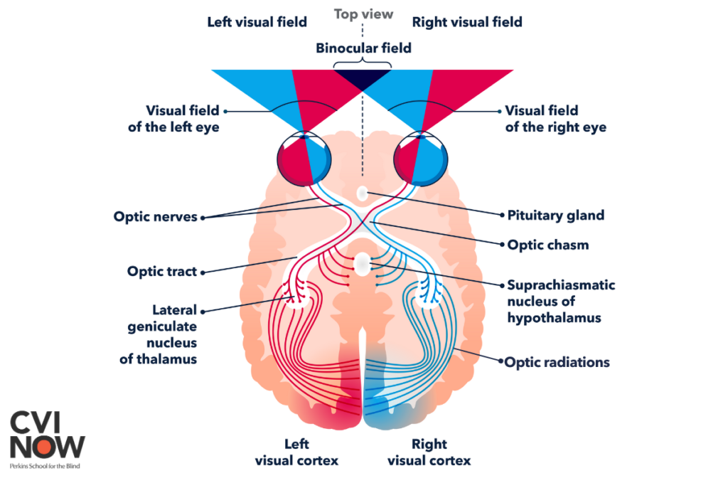
Obviously, from the lens to the visual cortex, there are many stops along the visual pathway. As with other multi-component systems, any disruption along the pathway can lead to visual impairments or even blindness. Since this article is not about the causes of blindness, I’ll not go into the details of these disruptions. Interested readers may visit dedicated organizations to learn more. For example, Perkins School of the Blind, or the National Federation of the Blind.
The point is, it’s more likely for blindness to result from malfunctions along this visual pathway than from an illness in the brain. Thus, most blind individuals’ brains are as healthy and as functional as those of sighted individuals. The brains of blind individuals contain all the necessary gadgets understand words and meanings. It’s just that blind individuals can’t take in the words through their eyes. Instead, blind individuals read with their fingers, using a spelling system called “braille”.
Unified English Braille (UEB)
Whereas we spell English words with letters, we spell braille words with “cells”. Across languages, most of the modern braille spelling systems agree upon a 6-dot cell. In a 6-dot braille cell, the dots form a 3-by-2 array. Conventionally, we label the 6 dots in the following order:

Each dot can either be raised above the surface on which the braille cell is printed, or stay flat. Since there are 2 possible configurations for each of the 6 dots, there are a total of 64 possible braille cells. Excluding the blank cell where all dots are flat, we have 63 of them. For most languages with an alphabetical spelling system, 63 cells are more than enough to represent all the letters in the alphabet, although the correspondence between cells and letters may be different across languages. For instance, when you see the following braille word:

While it means “apple” in English braille, it may mean something totally different in Polish braille. Just like the word “chat” has completely different meaning in English and French. Meanwhile, in French braille, these same five cells do correspond to the letters “a,p,p,l,e”, but there’s no such a word in French.
The form of Braille most commonly used by English-speaking proficient blind readers is called Unified English braille (UEB). Here are how the 26 letters are represented in UEB:
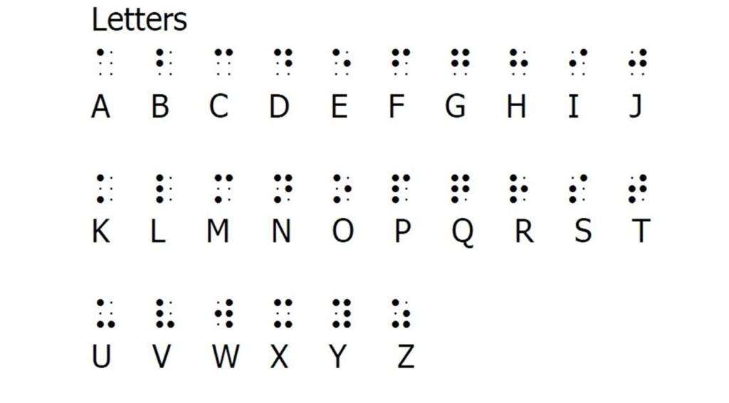
The use of “contractions”
To read braille, we have to swipe our fingers across the raised dots. It is the subtle tactile sensation of the dots gliding across our fingertips, rather than the pressure they apply to our skin, that enables the identification of braille cells. That is, even for fluent braille readers, it could be difficult to identify a cell by pressing a finger against it.
Imaginably, the size of our fingers constrains the size of the braille cells. We can’t enlarge or shrink braille cells, regardless of the surfaces on which we print them. We can’t squeeze the cells (too much), either. Braille cells should be properly spaced such that readers don’t mistakenly read the right column of the left cell and the left column of the right cell together as a cell. Due to these physical constraints, it takes much more space to spell English words in braille cells letter-by-letter. Here’s an example:

Moreover, for a braille writer, it could be effortful to emboss all the dots, especially for some very frequent words or letter combinations, such as -er or -ing. Out of such concerns, the designers of UEB incorporated a system of “contractions“. Recall that there are a total of 63 possible braille cells, and we only have 26 letters in the alphabet. It means that we have 37 spare cells to do some tricks. The basic idea is to use one or more braille cells to represent some Roman letter combinations. Of course, the number of braille cells in a contraction is always less than the number of Roman letters it represents.
Types of contractions
There are nuances in the terminologies and precise classifications of the types of contractions. However, we can roughly identify four types of contractions.
One cell to represent a group of letters or a word
- The uncontracted braille spelling of “ing“ is ⠊⠝⠛, but we can replace it with ⠬ . Similarly, “er(⠑⠗)” can be replaced with ⠻ . The cell used to represent a group of letters contained within a word are called “groupsign“. Other groupsigns include “ch(⠡)”, “st(⠌)”, “wh(⠱)”, “ar(⠜)”, “dis(⠲)”, “con(⠒)”, “en(⠢)”, and more.
- Some contracted groups of letters make up a word. In this case, we can use the contraction cell, standing alone, to represent that word. In the rulebook of UEB, such contractions are distributed across two categories, “strong contractions” and “lower wordsigns“. There are only 7 of them. They are: “and(⠯)”, “for(⠿)”, “of(⠷)”, “the(⠮)”, “with(⠾)”, “be(⠆)”, “in(⠔)”. We can use these contractions to represent the same group of letters within a word. For example, “without” is spelled as with-ou-t (⠾⠳⠞).
One stand-alone cell representing a whole word
- Most cells which stand for a single letter can stand for a whole word when used alone. For example, ⠓ stands for the letter “h” when it is part of a word like “happy” (⠓⠁⠏⠏⠽), but when presented alone, it stands for the word “have”. Other examples include “E(⠑)=every”, “K(⠅)=knowledge”, “P(⠏)=people”. We call such contractions “alphabetic wordsigns“.
- Some groupsigns also represent a whole word when standing alone. For example, “ch(⠡)=child”, “st(⠌)=still”, “wh(⠱)=which”, “en(⠢)=enough”. We call them “wordsigns” when they represent a whole words. “child”, “still”, and “which” are examples of “strong wordsigns“, whereas “enough” is an example of “lower wordsigns“. Here we don’t have to care about the distinction between “strong” and “lower”, though.
- As mentioned previously, “and”, “for”, “of”, “the”, “with”, “be”, and “in” can be both a wordsign or a groupsign.
Two cells to represent a group of letters
There are 5 cells which don’t stand for any letter or group of letter by themselves. Rather, we use them as “indicators” to modify the way we represent the cell that follows. These 5 indicator cells are: ⠘(dots 4, 5), ⠸(dots 4, 5, 6), ⠐(dot 5), ⠨(dots 4,6), ⠰(dots 5, 6)
- Initial-letter contractions.
Add [dots 4, 5], [dots 4, 5, 6], or [dot 5] in front of a cell to represent a word that begins with that cell. For example:- [dots 4, 5] + “u” (⠘⠥) = upon
- [dots 4, 5] + “th” (⠘⠹) = those
- [dots 4, 5] + “the” (⠘⠮) = these
- [dots 4, 5, 6] + “s” (⠸⠎) = spirit
- [dots 4, 5, 6] + “m” (⠸⠍) = many
- [dots 4, 5, 6] + “the” (⠸⠮) = their
- [dot 5] + “d” (⠐⠙) = day
- [dot 5] + “q” (⠐⠟) = question
- [dot 5] + “the” (⠐⠮) = there
- We can use an initial-letter contraction within a word to represent those letters. For example, “Germany (⠠⠛⠻⠸⠍)” = [indicator for capitalization: dot 6]+g+er+many. There are some complicated rules stipulating when we should not do this. Interested readers may refer to the rulebook of UEB.
- Final-letter groupsigns.
Add [dots 4, 6] or [dot 5, 6] in front of a cell to represent a group of letters that ends with that cell. For example:- [dots 4, 6] + “d” (⠨⠙) = -ound
- [dots 4, 6] + “s” (⠨⠎) = -less
- [dots 5, 6] + “n” (⠰⠝) = -tion
- [dots 5, 6] + “t” (⠰⠞) = -ment
- We use these groupsigns when the letters they represent come after other letters or contractions. For example, “rational” is spelled as ⠗⠁⠰⠝⠁⠇(r-a-tion-a-l). However, we don’t spell “mentor” as ment-o-r, because in this word, “ment” doesn’t come after any other letter. Instead, we spell “mentor” as ⠍⠢⠞⠕⠗ (m-en-t-o-r).
Shortforms
For some English words, we only have to write some of its letters to represent the whole word. For example: almost = ⠁⠇⠍ (a-l-m); braille = ⠃⠗⠇ (b-r-l); immediate = ⠊⠍⠍ (i-m-m); such = ⠎⠡(s-ch); because = ⠆⠉ (be-c). In some occasions, we can use a shortform to represent the corresponding group of letters in a longer word. However, there are again complicated rules stipulate when we can and can’t do this.
Basically, these are all we have to know about contractions as non-readers. If you spell out all English words using only the 26 alphabetical cells, we say you’re using “grade 1” UEB. If you follow the rules of contractions and use them properly, you’re using “grade 2” UEB. Most braille readers learned grade 1 spellings at first, then advanced to grade 2 contractions.
To understand my braille reading project, we only have to know this much about braille. The next piece of necessary background knowledge is the neural basis of sighted reading.
The Visual Word Form Area (VWFA) for visual reading
Knowing the drastic difference between visual and tactile writing systems, we may appreciate the hypothesis that visual and tactile reading involve different brain mechanisms. Then, what could the difference be?
In visual reading, one particular important brain region is the Visual Word Form Area (VWFA). This region is a part of the left ventral occipito-temporal cortex (vOTC) – on the bottom surface of our left brain, close to the back of our head. The VWFA plays a crucial role in the recognition of written language, particularly words and letter strings.
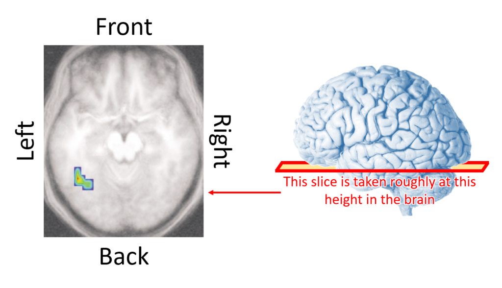
Left figure credit: McCandliss et al., 2003
Right image credit: https://www.purposegames.com/game/lateral-view-of-the-brain-quiz-game
Interestingly, in the brain of a newborn or an illiterate adult, the left vOTC does not respond preferentially to written words. The vOTC develops into the VWFA – visual word form area – only after the acquisition of literacy. Across languages and the ages of literacy acquisition, it’s always the same region in the left vOTC which eventually becomes the VWFA. Why?
One popular hypothesis is that the left vOTC lies at the confluence of visual and linguistic inputs. Anatomically, the vOTC is connected with both the primary visual cortex in the occipital lobe and the language processing regions in the frontal lobe. When we learn how to read, we explicitly associate the visual form of a word and its linguistic properties. According to this hypothesis, this association leads to the vOTC accepting information input from both the visual and the language regions. Eventually, it develops into a region bridging the visual word form of a word and its linguistic contents – the VWFA.
If the origin story of the VWFA is true, then what about people who have never received visual input during the acquisition of literacy?
Could a “tactile” word form area lie elsewhere in the brain?
Congenitally blind (CB) individuals – people who have been blind since birth – never learned how to read visually. Such individuals have always been reading with their fingertips. Therefore, we shouldn’t expect the word form area of CB readers to develop at the confluence of visual and language information. To begin with, the occipital “visual” cortex of CB individuals has never processed any visual information.
On the contrary, we could expect the word form area for CB individuals to develop at the confluence of language and tactile information. One candidate for such a “tactile” word form area is the supramarginal gyrus (SMG) in the posterior parietal cortex (PPC). It’s about 5cm (2 inches) above your ears.
Here’s a schematics for our hypothesis regarding how a visual and a tactile word form area can develop in different parts in the brain.
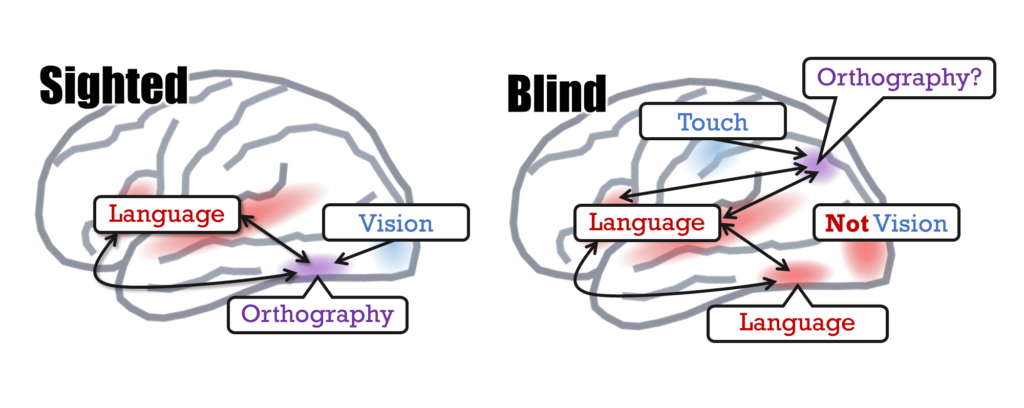
In the figure, we use the word “orthography“, which we can roughly understand as “how a word is spelled“. This is one of the crucial properties a purported word form area cares about. Conversely, we can say that caring about orthography is a signature of a word form area. Therefore, in our study, we ask:
Which region(s) in the brain of congenitally blind individuals processes braille orthography?
To find out, we took advantage of the system of contraction in United English Braille (UEB). Throughout the life of a sighted English reader, they only learn one system of orthography: the Roman spellings of English words. There’s only one way to spell a word like “teacher”. On the contrary, congenitally blind braille readers learned two systems of orthography early in life: uncontracted (grade 1) and contracted (grade 2) UEB. To CB readers, the word “teacher” can be spelled in two remarkably different ways: ⠞⠑⠁⠉⠓⠑⠗(t-e-a-c-h-e-r) and ⠞⠂⠡⠻(t-ea-ch-er). Even as an adult, CB readers are fully aware of the two sets of orthography.
Hence, the specific goal of our research project is to identify the brain region(s) which juggles the contracted and uncontracted spelling systems.
Same word length when contracted, different when uncontracted
In our experiment, congenitally blind braille readers read braille words while undergoing functional magnetic resonance imaging (fMRI) scans. As you might know, MRI scanners produce super strong magnetic fields which interferes with normal electronic devices, including mechanical braille displays. Fortunately, Dr. Merabet Lotfi kindly lend us an MRI-compatible braille display. The manufacture of this unique braille display is another epic worth recounting. But, let me just save it in my bucket-list of site contents, and focus on our experiment for the moment.
Throughout the two-hour long scanning session, participants read the same set of 30 words for 30 times. The order of the words was randomized in each repetition. Here are the words:
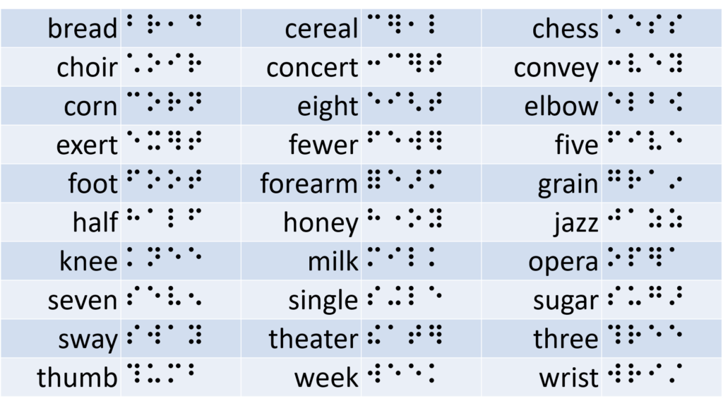
Did you notice a feature common across the words? All these words can be spelled with exactly 4 braille cells, thanks to the existence of contractions. On the contrary, their uncontracted spellings range from 4 to 7 letters/cells.
Most congenital blind braille readers first learned the grade 1 (uncontracted) spellings, and later progressed to grade 2 spellings with contractions. The braille information plates (such as the ones on ATM) blind readers encounter every day are written in contracted spellings. Meanwhile, when communicating with sighted individuals, such as talking to some customer service personnels, blind people can spell out words in the uncontracted form. Therefore, braille readers are well aware of the two spelling systems – the two systems of braille orthography.
The “supramarginal gyrus” is sensitive to the difference between the two systems of braille orthography
Through fMRI scanning, we recorded the neural response associated with each of the 30 words. Then, we asked which region(s) in the brain showed neural activity proportional to the uncontracted length of the word.
Here’s the rationale for this question. All the words participants read consisted of exactly 4 cells. So, the neural response to the physical length of the words should be the same. We can’t derive the uncontracted word length from the physical properties of the words. To know how many letters/cells there should be in the uncontracted spelling in a word, we have to understand both the contracted and uncontracted orthography, and mentally convert the two systems. That is, given the same physical lengths of the words, if the neural response in a region is proportional to the uncontracted word length, such a region is likely to underlie said orthographic conversion.
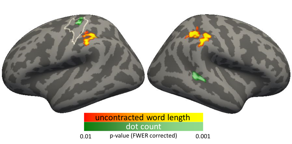
We indeed identified such a region. It is the “supramarginal gyrus (SMG)” in both halves of the brain (red/orange in the following figure). This region is close to the primary somatosensory (S1) cortex, where the brain processes tactile input. Perhaps the supramarginal gyrus is to the S1 what the ventral occipito-temporal cortex is to the primary visual cortex. Whereas the vicinity to visual input transformed the ventral occipital-temporal cortex into the visual word form area, the vicinity to the somatosensory cortex may have transformed the supramarginal gyrus into the tactile word form area.
Control for the amount of physical stimulation
However, before we draw any conclusion, we have to make sure the response in the SMG didn’t result from physical stimulation. On average, cells for contractions use one more dot than non-contraction cells. Thus, there remains the possibility that the SMG activity is proportional to the number of dots in a word. We considered this possibility in our analysis. We found that was not the case.
The region where the neural response was proportional to the number of dots was the S1 in the left brain. Not the SMG. Therefore, the SMG response is indeed proportional to the uncontracted word length, independent of the amount of physical stimulation (dot count).
Another evidence: SMG shows greater response to pronounceable non-words than real words
During the experiment, besides the 30 words, participants also encountered 30 “pseudowords”. A pseudoword is a pronounceable non-word. In the brains of sighted readers, one property of the VWFA is a stronger response to pseudowords than to real words.
Using our data collected from blind readers, when we contrasted pseudowords against real words, the SMG was once again among the regions showing stronger response.
Therefore, the SMG may be responsible for the conversion between different braille orthographic systems, and it responds more strongly to pseudowords than to real words. Both properties support the hypothesis that the SMG may be the tactile word form area we’re looking for. However, is it really so? Our current findings are reported in the Journal of Cognitive Neuroscience. But it is only the first step towards a series of future investigations. There is still much work to do before we can say for sure that we found the braille-reading counterpart of the visual word form area.
References
- Ackland, P., Resnikoff, S., & Bourne, R. (2017). World blindness and visual impairment: despite many successes, the problem is growing. Community Eye Health, 30(100), 71-73.
- Simpson, C. (2013). The rules of unified English braille (2nd ed.): International Council on English Braille.
- Liu, Y.-F., Rapp, B., & Bedny, M. (2023). Reading Braille by Touch Recruits Posterior Parietal Cortex. Journal of Cognitive Neuroscience, 1-24. doi:10.1162/jocn_a_02041
- Millar, S. (1997). Reading by touch: Routledge.
- McCandliss, B. D., Cohen, L., & Dehaene, S. (2003). The visual word form area: expertise for reading in the fusiform gyrus. Trends in Cognitive Sciences, 7(7), 293-299. doi:https://doi.org/10.1016/S1364-6613(03)00134-7

